 Cellpose-napari + FIJI#
By Frederic Bonnet 🔬
Learning Objectives#
In this lesson, you’ll learn how to use the Cellpose napari plugin in conjunction with FIJI to complete a segmentation workflow.
Time to learn: 1 hour
Prerequisites#
Napari version and plugins |
Importance |
Notes |
|---|---|---|
Mandatory |
In the napari viewer, verify your version of napari by clicking on the Help menu, then napari info. |
|
Mandatory |
Install this plugin from within the napari viewer, by going to the Plugin menu, then clicking on Install/uninstall plugins. Search for Cellpose-napari and click install. |
|
Mandatory |
||
Mandatory |
Allows us to open our image’s mask in FIJI. |
|
tiffs, JPEGs or PNGs |
Mandatory |
Any of these three file types will be supported |
Single plane images |
Optional |
Channels can be read as: (nY x nX x) or (x nY x nX) |
Multiplane images |
Optional |
Required image shape: (nZ x nY x nX) |
Multichannel images, like multi-Z tiffs |
Optional |
The expected format: (nZ x nC x nY x nX) |
Optional |
Because this is 2D data, only 2D segmentation may be applied to it. |
What does this plugin do?#
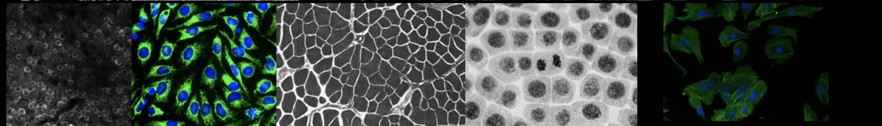
Cellpose-napari utilizes a generalist algorithm for cell and nucleus segmentation. It allows you to perform segmentation workflows on cell bodies, membranes and nuclei in images. It also has deep-learning built-in, allowing it to segment many types of cells without requiring parameter adjustments, new training data, or further model retraining. Because of its versatility, it supports a wide variety of microscopy modalities and fluorescent markers.
Demo of Cellpose-napari#
Here are a few recommendations before you watch the video:
Use full screen view
Slow the video down by clicking the gear wheel or cog wheel in the lower right corner of the video. Click speed and select a slower speed.
Review the parameter setting explanations here. In the video the following parameters and features are used:
In napari:
contrast limits
loading a plugin
In the cellpose-napari plugin:
model type
channel to segment
diameter
computer diameter from image
average 4 nets
resample dynamics
clear previous results
output flows and cellprob
output outlines
run segmentation
cellprob threshold
recompute last masks with new cellprob + model match
Hint
When any of the buttons are clicked, it may take a few seconds for napari to respond with the new information. Please be patient.
The above video demonstrates how to open the Cellpose-napari plugin, segment your image, and adjust its parameters. If you would like a more detailed explanation of each parameter, proceed to the next part of this lesson.
Note
Because napari can’t export an image’s mask at this time, we must use FIJI for this. The final part of this lesson covers segmenting your image with cellpose-napari, then using FIJI to export and analyze your image’s mask.